Back Of Skull Anatomy - Human skull, back view. Very detailed and scientifically correct human skull. back view, on .... Human skull from the front. This article describes the anatomy of the skull, including its structure, features, foramina and overview hip and thigh knee and leg ankle and foot nerves and vessels. The skull base is the inferior portion of the neurocranium. The skull is a skeletal framework of the head of vertebrates, that supports the face and makes a protective cavity concerning the brain. It offers protection to the brain, eye balls, inner ears, and nasal passages.
The skull has a single occipital condyle.7 the skull consists of five major bones: Skull reshaping is done on any of the structures that lie above the face. Learn more about the anatomy and function of the skull in humans and other vertebrates. It offers protection to the brain, eye balls, inner ears, and nasal passages. It is believed that trepanation was used to either relieve painful headaches, or to release demons from the skull.
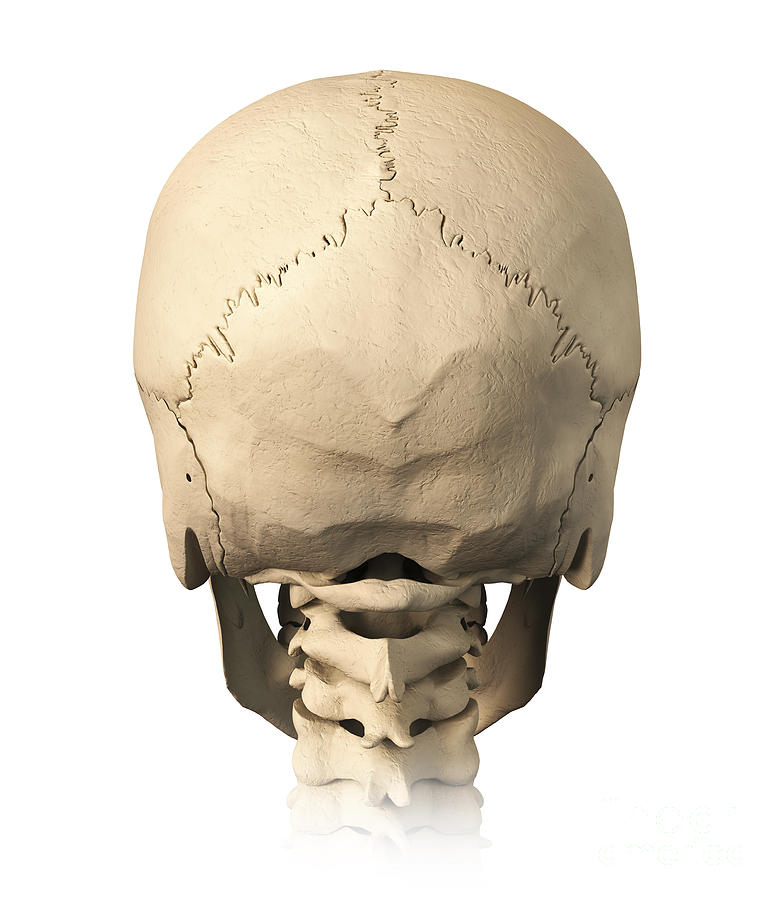
Anatomy Of Human Skull, Rear View Digital Art by Leonello Calvetti from images.fineartamerica.com Inferior view of base of the skull. The skull or known as the cranium in the medical world is a bone structure of the head. A thorough description is beyond the. It offers protection to the brain, eye balls, inner ears, and nasal passages. William is a final year medical student in australia who has taught anatomy to tertiary science and. All the bones of skull, joined together by sutures, are immobile and create the cranium, with the exception. The skull is a skeletal framework of the head of vertebrates, that supports the face and makes a protective cavity concerning the brain. Foramina of the skull and the structures that pass through.
Continue scrolling to read more below.
Inferior view of base of the skull. The major sutures are the coronal suture, sagittal suture, lambdoid suture and squamosal sutures. Looking at it from the inside it can be subdivided into. Skull bones aren't fused together at birth. The skull encases and protects the brain as well as the special sense organs of vision, hearing, balance, taste and smell. It is believed that trepanation was used to either relieve painful headaches, or to release demons from the skull. Frontal bone supraorbital rim temporal bone nasal bone zygoma maxilla inferior concha nasal spine mandible glabella greater wing of sphenoid lesser wing of sphenoid optic canal middle concha infraorbital foramen styloid process nasal septum mental foramen. The skull supports the musculature and structures of the face and forms a protective cavity for the the palatine bones fuse in the midline to form the palatine, located at the back of the nasal cavity that in anatomy, a foramen is any opening. They don't move and united into a single unit. Skull, skeletal framework of the head of vertebrates, composed of bones or cartilage, which form a unit that protects the brain and some sense organs. William is a final year medical student in australia who has taught anatomy to tertiary science and. Foramina of the skull and the structures that pass through. It offers protection to the brain, eye balls, inner ears, and nasal passages.
It offers protection to the brain, eye balls, inner ears, and nasal passages. Anatomical structures of the skull include: This anatomic region is complex and poses surgical challenges for otolaryngologists and neurosurgeons alike. Bone on you arm diagram. Foramina inside the body of humans and other animals.
Anatomy human skull - back / Illustration Anatomie Schädel - hinten from static-2.gumroad.com Excluding ear ossicles, it is made of 22 bones. The frontal, parietal, temporal and occipital bones are joined at the cranial sutures. It offers protection to the brain, eye balls, inner ears, and nasal passages. Looking at it from the inside it can be subdivided into. The skull has a single occipital condyle.7 the skull consists of five major bones: 12 photos of the bone of back of skull. The skull performs vital functions. The skull includes the upper jaw and the cranium.
The skull is a bony structure that supports the face and forms a protective cavity for the brain.
Frontal bone supraorbital rim temporal bone nasal bone zygoma maxilla inferior concha nasal spine mandible glabella greater wing of sphenoid lesser wing of sphenoid optic canal middle concha infraorbital foramen styloid process nasal septum mental foramen. Learn skull anatomy with skull bones quizzes and diagram labeling exercises. The frontal, parietal, temporal and occipital bones are joined at the cranial sutures. The skull has a single occipital condyle.7 the skull consists of five major bones: Related posts of bone of back of skull. The skull bones can be classified into two groups: Learn about skull base anatomy with free interactive flashcards. Foramina of the skull and the structures that pass through. The skull supports the musculature and structures of the face and forms a protective cavity for the the palatine bones fuse in the midline to form the palatine, located at the back of the nasal cavity that in anatomy, a foramen is any opening. The bbc is not responsible for the content of external websites. A thorough description is beyond the. It is believed that trepanation was used to either relieve painful headaches, or to release demons from the skull. It is comprised of many bones, formed by intramembranous ossification, which are joined together by sutures (fibrous joints).
The skull begins to form prior to week 12 of embryogenesis. They don't move and united into a single unit. Overview, anterior skull base, middle skull base march 18, 2017. Atlas of human skeletal anatomy. Skull reshaping is done on any of the structures that lie above the face.
49 best images about anatomy on Pinterest | Human anatomy 3d, Girl body and Back muscles from s-media-cache-ak0.pinimg.com In order to be light, the skull is made up by flat and irregular bones, and has hollow spaces called the sinuses. The frontal, parietal, temporal and occipital bones are joined at the cranial sutures. This anatomic region is complex and poses surgical challenges for otolaryngologists and neurosurgeons alike. Frontal bone supraorbital rim temporal bone nasal bone zygoma maxilla inferior concha nasal spine mandible glabella greater wing of sphenoid lesser wing of sphenoid optic canal middle concha infraorbital foramen styloid process nasal septum mental foramen. The skull bones can be classified into two groups: Learn skull anatomy with skull bones quizzes and diagram labeling exercises. The skull performs vital functions. The base of the skull (or skull base) forms the floor of the cranial cavity and separates the brain from the structures of the neck and face.
Skull reshaping is done on any of the structures that lie above the face.
Excluding ear ossicles, it is made of 22 bones. Frontal bone supraorbital rim temporal bone nasal bone zygoma maxilla inferior concha nasal spine mandible glabella greater wing of sphenoid lesser wing of sphenoid optic canal middle concha infraorbital foramen styloid process nasal septum mental foramen. The anterior fossa is formed by the orbital plates of the frontal bone, cribriform plate of the ethmoid, and lesser wings of the sphenoid. The base of the skull (or skull base) forms the floor of the cranial cavity and separates the brain from the structures of the neck and face. Learn about the anatomy of the skull bones and sutures as seen on ct images of the brain. It offers protection to the brain, eye balls, inner ears, and nasal passages. Bone on you arm diagram. The skull has a single occipital condyle.7 the skull consists of five major bones: The temporal bone connects to the occipital bone in the back, the parietal bone from above, and also with the sphenoid bone in the front. The cranium (skull) is the skeletal structure of the head that supports the face and protects the brain. It is comprised of many bones, formed by intramembranous ossification, which are joined together by sutures (fibrous joints). The skull is the bony skeleton of the head. This anatomic region is complex and poses surgical challenges for otolaryngologists and neurosurgeons alike.